Myofibrils
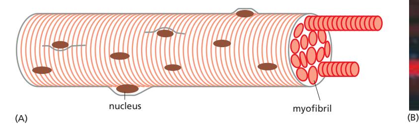 Fig 16–28. 골격근 세포의 구조. 근섬유(muscle fiber)는 여러 myofibril로 구성되고, 각 myofibril은 직렬로 배열된 sarcomere로 이루어진다
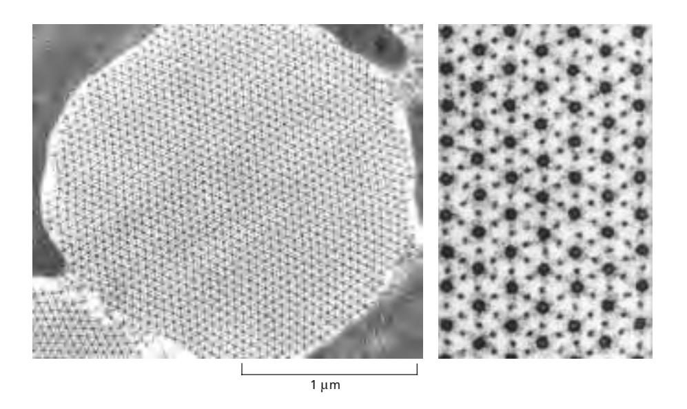 insect flight muscle의 cross section.
acitn과 myosin filaments가 pack되어있음. 수백 수천개의 sarcomere가 보인다.
커다란 단면 하나가 1 myofibril. 직경 1~2μm
insect flight muscle의 cross section.
acitn과 myosin filaments가 pack되어있음. 수백 수천개의 sarcomere가 보인다.
커다란 단면 하나가 1 myofibril. 직경 1~2μm
구조
골격근 세포는 myofibril이라 불리는 긴 수축성 구조물들로 채워져 있다. 각 myofibril은 수천 개의 sarcomere가 직렬로 연결된 구조이며, sarcomere는 actin thin filament와 myosin thick filament가 고도로 정렬된 최소 수축 단위이다. 길이 : 2μm
전자현미경으로 보면 myofibril에 줄무늬가 보이는데, 이는 sarcomere의 반복적 배열에서 비롯된다:
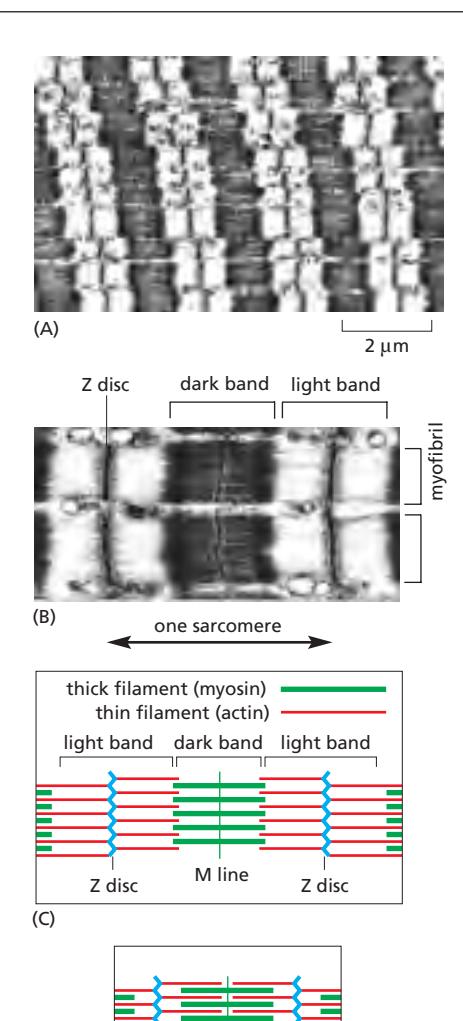
- Z disc (Z line): Sarcomere의 양 경계를 이루며, α-actinin을 포함. Actin 필라멘트의 plus end(barbed end)가 여기에 고정된다.
- Light band(I band): Z disc 양쪽에 위치하며 actin thin filament만 포함 → 밝게 보임.
- Dark band(A band): Myosin thick filament 전 구간 → 어둡게 보임. Actin 필라멘트가 겹치는 구간 포함.
- H zone: A band 중앙으로 myosin만 있고 actin이 겹치지 않는 구간.
- M line: Thick filament 중앙에 위치하며 myosin tail을 연결하는 단백질로 구성.
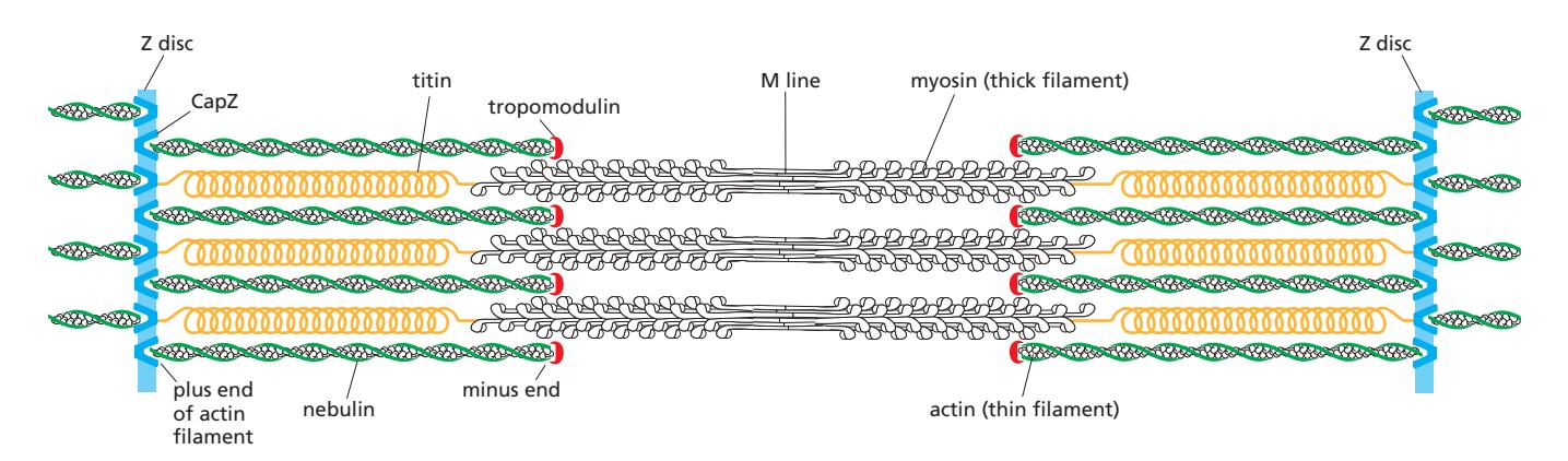
accessory proteins in sarcomere
thin filaments의 길이를 일정하게 하는 proteins
CapZ
Z disc에 thin filaments의 plus end를 anchoring하고 성장과 분해를 막음.
tropomodulin
thin filaments의 minus end에 붙어 성장과 분해를 막음. CapZ와 tropomodulin에 의해 light band의 actin filaments가
nebulin
thin filament와 동일한 길이의 단백질로 나선형으로 thin filament 측면에 붙어 안정화함.
myosin에 관여하는 protein
titin
스프링 형태로, myosin thick filaments와 Z disc를 연결함. sarcomere에 탄성을 부여
tropomyosin
thin filaments를 따라 측면에 나선형으로 부착되어 있음. Ca이온 농도가 낮을 때 actin과 myosin의 결합부위를 막는 단백질.
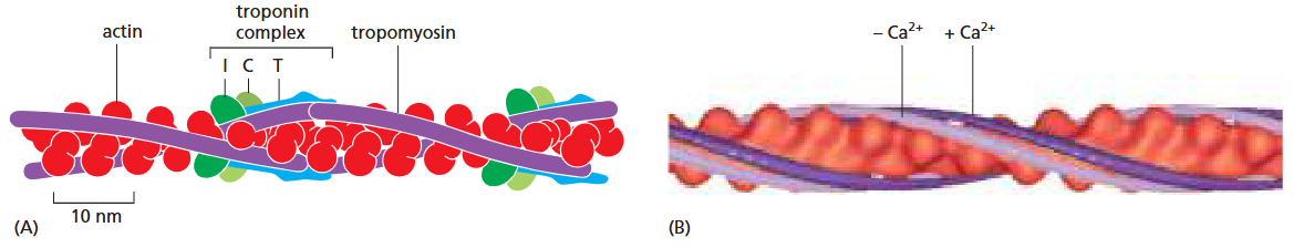
troponin
troponin complex로 존재하며, Tn I, Tn C, Tn T subunit으로 구성됨. Ca이온 농도에 따라 tropomyosin의 위치를 변경하여 myosin과 actin의 결합부위 노출을 조절함. 각각의 역할은 troponin참조.
Sliding Filament Model
근육 수축 시 sarcomere 길이가 줄어든다. 이는 actin thin filament와 myosin thick filament 자체의 길이 변화 없이, 두 필라멘트가 서로를 향해 미끄러지기 때문이다. Myosin head의 반복적 power stroke가 actin 필라멘트를 M line 쪽으로 잡아당겨 Z disc 간의 거리를 좁힌다.
accessory protein 배치는 Organization of accessory proteins in a sarcomere, Ca²⁺ 신호 전달은 T tubules, sarcoplasmic reticulum, troponin 참조.