Structured Illumination Microscopy (SIM)
개요
회절 한계를 극복하는 첫 번째 superresolution 접근법으로, 약 100 nm의 resolution을 가진 fluorescence imaging 방법이다. 이는 conventional bright-field microscopy의 약 2배 resolution에 해당한다.
회절 한계의 극복
Light microscopy의 변형들은 모두 classic diffraction limit에 의해 제약받아 약 0.2 μm(200 nm)까지만 분해능을 가진다(Figure 9-5 참조). 그러나 여러 접근법들이 빛의 회절로 인한 한계를 우회하며, 일부는 10 nm 정도의 작은 객체까지 성공적으로 분해할 수 있어 20배의 놀라운 개선을 이뤘다.
SIM의 원리
기본 개념
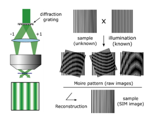
SIM은 grated 또는 structured pattern의 빛으로 sample을 조명하여 회절 한계를 극복한다. Microscope의 물리적 설정과 작동은 상당히 복잡하지만, 일반적인 원리는 moiré pattern(모아레 패턴) 생성과 유사하게 생각할 수 있다.
Moiré Pattern(모아레 패턴)
Moiré pattern은 서로 다른 각도나 mesh 크기를 가진 두 grids를 겹쳐 만든 interference pattern이다. 조명 grid와 sample features가 결합되어 interference pattern을 만들며, 여기서 grid spacing보다 작은 features가 더 큰 patterns로 변환된다.
Figure 9-27: Structured Illumination Microscopy 원리

(A) 미지의 구조로부터의 pattern: 원래 구조의 pattern을 보여줌 (B) 정의된 grid pattern: 알려진 grid pattern (C) Moiré pattern: 이 둘을 결합하면 resulting moiré pattern이 A(원래 pattern)에서 쉽게 보이는 것보다 더 많은 정보를 포함함
원리: 알려진 pattern(B)이 더 높은 spatial frequencies를 가지면 더 나은 resolution을 얻을 수 있음. 그러나 광학적으로 생성할 수 있는 spatial patterns도 회절 제한을 받기 때문에, SIM은 resolution을 약 2배 정도만 개선할 수 있다.
작동 메커니즘
Image Acquisition
Classical limit을 넘어서는 원래 features가 optical system에 의해 imaging될 수 있도록 변환됨. Computer image processing은 이들을 classical limit의 2배까지 resolution을 가진 이미지로 복원할 수 있다.
Grid Illumination 과정
문제: Grid의 dark stripes에 있는 sample 부분은 조명되지 않아 imaging되지 않음 해결: Imaging을 여러 번(일반적으로 3번) 반복. 각 이미지 사이에 grid를 grid spacing의 일부만큼 이동시킴
방향성 고려: Interference effect는 grid bars 방향에 가까운 image components에서 가장 강함. 따라서 모든 방향에서 동등한 enhancement를 얻기 위해 전체 과정을 grid pattern을 일련의 각도로 회전시키며 반복함.
최종 단계: 이 모든 개별 이미지들을 computer로 수학적으로 결합하여 enhanced superresolution image 생성
SIM의 특징
다양성 (Versatility)
주요 장점: 어떤 fluorescent dye나 protein과도 사용 가능 3D 데이터: 연속적인 focal planes에서 캡처한 SIM images를 결합하여 three-dimensional data sets 생성 가능
Resolution
개선 정도: Conventional light microscopy 대비 약 2배 향상 한계: 광학적으로 생성 가능한 spatial patterns도 회절 제한을 받음
Figure 9-28: SIM을 이용한 3D 데이터 생성
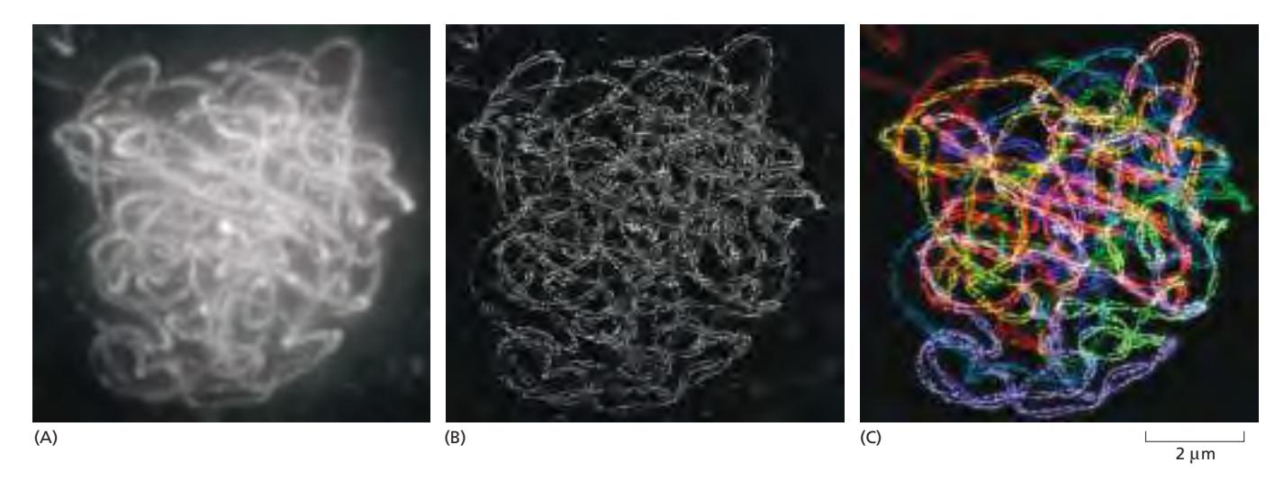
표본: Maize cell의 pachytene 단계 meiotic chromosomes의 three-dimensional projections 염색: Synaptonemal complexes의 paired lateral elements를 보여줌
(A) Conventional Fluorescence Microscopy: Chromosome set이 cohesin에 대한 fluorescent antibody로 염색됨. 두 lateral elements 사이의 거리가 약 200 nm(회절 한계)이기 때문에 각 complex를 구성하는 두 lateral elements가 분해되지 않음.
(B) 3D SIM Image: 향상된 resolution으로 각 lateral element(약 100 nm 폭)를 명확히 분해할 수 있으며, 각 개별 쌍의 두 chromosomes가 서로 coil하는 것을 볼 수 있음.
(C) 색상 추적: 전체 nucleus에 대한 complete three-dimensional data set이 가능하므로, 각 개별 chromosome 쌍의 경로를 추적하고 인위적으로 다른 색상을 할당할 수 있음.
SIM의 장점
- 광범위한 호환성: 모든 fluorescent dyes와 proteins 사용 가능
- 3D Imaging: Three-dimensional data sets 생성 가능
- 상대적으로 간단: 다른 superresolution 기술에 비해 구현이 용이
- 살아있는 세포: Living cells에서 사용 가능
- 빠른 Imaging: 비교적 빠른 acquisition time
SIM의 한계
- 제한된 Resolution 향상: 약 2배만 개선 (200 nm → 100 nm)
- 복잡한 Image Processing: 여러 이미지를 수학적으로 결합해야 함
- 광표백: Multiple acquisitions로 인한 photobleaching 증가
- Artifacts 가능성: Image processing 과정에서 artifacts 발생 가능
다른 Superresolution 기술과 비교
vs Confocal Microscope: Confocal은 회절 한계 내(200 nm), SIM은 약 100 nm resolution vs STED: STED는 약 20 nm resolution 가능하지만 특수한 photoswitchable probes 필요 vs PALM/STORM: 이들도 약 20 nm resolution이지만 더 긴 acquisition time과 특수 probes 필요
응용 분야
Chromosome Structure: Meiotic chromosomes의 synaptonemal complexes (Figure 9-28) Cell Biology: 회절 한계보다 작지만 10 nm까지는 아닌 구조들 Live Cell Imaging: 다른 superresolution 기술보다 빠른 imaging Tissue Imaging: 두꺼운 표본에서 3D reconstruction
기술적 고려사항
Grid Pattern: 적절한 spatial frequency 선택 각도와 위상: 여러 각도와 위상에서 imaging 필요 Computational Power: 복잡한 image reconstruction 알고리즘 Illumination 균일성: 균일한 grid pattern 조명 필요
요약
핵심 원리: Structured illumination과 moiré effect를 이용하여 회절 한계 극복, 약 2배 resolution 향상 주요 특징: 모든 fluorescent probes 사용 가능, 3D imaging 가능, 살아있는 세포에 적용 가능 Resolution: 약 100 nm (conventional의 2배) 응용: Chromosome structure, subcellular organelles, living cell dynamics 한계: 제한된 resolution 향상, 복잡한 image processing, photobleaching
참고 문헌
Chapter 9: Visualizing Cells and Their Molecules, Section: “Superresolution Fluorescence Techniques Can Overcome Diffraction-limited Resolution”, Related Figures: 9-27, 9-28, Related concepts: deconvolution, Confocal Microscope