Actin Arrays
세포 내 actin filament는 여러 accessory protein의 작용을 통해 다양한 형태의 array로 조직화된다. 주요 array는 세포의 피질(cortex)에서 만들어지며, 각각 특정 nucleating protein에 의해 개시된다.1
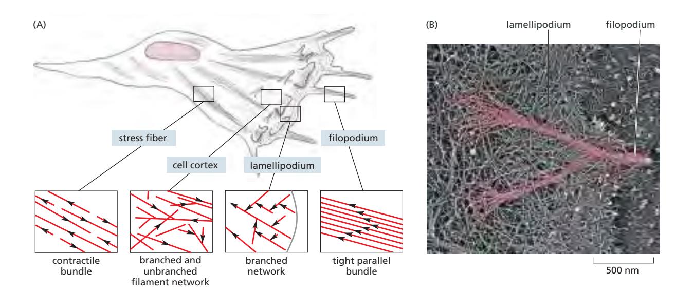 Fig 16–18. 세포 이동 중 fibroblast(섬유아세포. 이동성 좋음)의 actin array 모식도. (A) stress fiber(수축성), actin cortex(lamellipodia용 network), filopodia(spike형), lamellipodium. (B) Leading edge의 dense actin network. Plus end가 세포 끝을 향한다.
화살표가 향하는 방향이 minus end 쪽.
Fig 16–18. 세포 이동 중 fibroblast(섬유아세포. 이동성 좋음)의 actin array 모식도. (A) stress fiber(수축성), actin cortex(lamellipodia용 network), filopodia(spike형), lamellipodium. (B) Leading edge의 dense actin network. Plus end가 세포 끝을 향한다.
화살표가 향하는 방향이 minus end 쪽.
Rho GTPase Family에 의해 조절된다.
주요 Array 유형
dendritic network (lamellipodium)
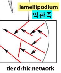
- Arp2/3 complex가 기존 filament 측면에서 새 filament를 약 70° 각도로 nucleate
- 2차원 sheet-like 구조로 세포 전진 방향에 평행하게 배열
- 방향성이 있음.
- 세포 가장자리를 밀어내는 힘 생성
- Rac GTPase에 의해 조절됨
- cell migration by actin cytoskeleton에 그 기전 설명됨.
Tight parallel bundle (filopodium)
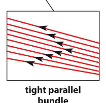
- Formin이 long, straight filament 형성
- 1차원 spike 구조, 세포 환경 탐색 및 신경 성장원뿔 유도 기능
- Plus end가 세포 끝을 향해 배치
- Cdc42 GTPase에 의해 조절됨
Contractile bundle (stress fiber)
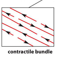
- 반대 극성의 actin filament가 myosin II와 함께 수축성 구조 형성
- Focal adhesion에 연결되어 세포가 기질에 traction 생성
- 번들을 형성하여 존재.
- 방향성은 양방향으로 존재.
- Myosin II가 수축력을 제공
- Rho GTPase에 의해 조절됨.
gel-like network(cell cortex)
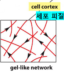
- Plasma membrane 바로 아래, 2D 또는 3D network
- 세포에 기계적 강도 제공, membrane shape 유지
- 명확한 방향성이 없음.
각 array 형성에 관여하는 단백질의 자세한 내용은 major accessory proteins of the actin cytoskeleton 참조.
Footnotes
-
2023 기말 16번 — Arp2/3에 의한 branched network와 formin에 의한 linear filament가 서로 다른 actin array를 구성한다는 내용이 정답 근거로 활용됨. ↩