Cortical Cytoskeleton
Cortical cytoskeleton은 대부분의 세포에서 plasma membrane 아래에 존재하는 specialized cytoskeletal network이다. 이는 membrane에 mechanical strength를 제공하고 Membrane protein의 확산을 제한하는 중요한 역할을 한다.
Red Blood Cell의 Spectrin-based Cytoskeleton
구조와 기능
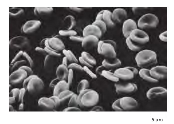
Human red blood cell의 특징적인 biconcave shape은:
- Plasma membrane protein과 underlying cytoskeleton 사이의 상호작용으로 인함
- Cytoskeleton은 주로 filamentous protein spectrin의 meshwork로 구성됨
미세혈관에서 접힐때도 있음. spectrin의 탄성으로 모양 복귀 가능
Red Blood Cell의 특수성:
- 세포의 유일한 membrane이 plasma membrane
- Nucleus와 다른 organelle 없음
Spectrin의 구조
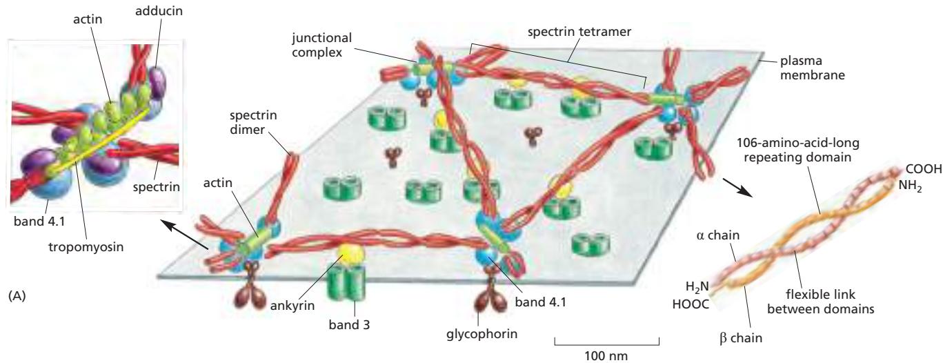
Spectrin:
- 길고 얇은 flexible rod
- 약 100 nm 길이
- Red blood cell cytoskeleton의 주요 성분
기능:
- Plasma membrane의 구조적 integrity와 shape 유지
- 세포가 좁은 capillary를 통해 강제될 때 membrane에 가해지는 stress 견딤
유전적 이상: Mice와 human에서 spectrin의 유전적 이상:
- Anemia 발생
- Red blood cell이:
- Spherical (concave 대신)
- Fragile
- Anemia의 심각도는 spectrin 결핍 정도에 증가
Nucleated Cell의 Cortical Cytoskeleton
구조적 특징
대부분의 다른 세포에서:
- Analogous하지만 훨씬 더 elaborate하고 highly dynamic한 cytoskeletal network 존재
- 세포의 cortex 구성
- Actin filament가 풍부함
- 다양한 방식으로 plasma membrane에 부착
Cortical cytoskeleton:
- Cell cortex의 일부
- Red cell cytoskeleton보다 복잡
- Spectrin과 red cell cytoskeleton의 다른 성분에 구조적으로 homologous한 protein 포함
동적 특성과 기능
Cortical actin network의 dynamic remodeling은:
- 많은 필수 세포 기능을 위한 driving force 제공
- Cell movement
- Endocytosis
- Transient, mobile plasma membrane structure 형성:
Membrane Protein Diffusion 제한
Corralling 메커니즘
Cortical cytoskeletal network은:
- 직접 anchor된 것 이상의 plasma membrane protein diffusion 제한
- Cytoskeletal filament가 plasma membrane의 cytosolic surface에 밀접하게 apposed
- Mechanical barrier 형성
- Membrane에서 protein의 자유 확산 방해
위 그림 (A)는 filament가 membrane을 작은 domain (corral)로 partition하는 diffusion barrier를 제공하는 것을 보여준다.
Corral의 특성:
- Permanent 또는 transient 가능
- Filament가 membrane과의 attachment를 일시적으로 잃으면 protein이 인접 corral로 escape 가능
Confinement 정도
위 그림 (B)는 high-speed single-particle tracking을 사용하여 fluorescently labeled membrane protein 하나의 경로를 시간에 따라 추적한 것을 보여준다:
- 개별 protein molecule이 tightly delimited membrane domain 내에서 확산
- 드물게 이웃 domain으로 escape(hop diffusion) (색 변화로 표시)
Transmembrane protein이 corral 내에 국한되는 정도는 다음에 의존:
- 다른 protein과의 association
- Cytoplasmic domain의 크기
큰 cytosolic domain을 가진 protein:
- Cytoskeletal barrier를 통과하기 더 어려움
예시:
- Cell-surface receptor가 extracellular signal molecule에 결합
- Large protein complex가 receptor의 cytosolic domain에 형성
- Receptor가 corral에서 escape하기 더 어려워짐
기능적 의의:
- Corralling이 signaling complex를 집중시키는데 도움
- Signaling process의 속도와 효율 증가