microtubule은 minus end 쪽(α-tubulin)보다 plus end 쪽(β-tubulin)에서 더 빠른 성장을 한다.
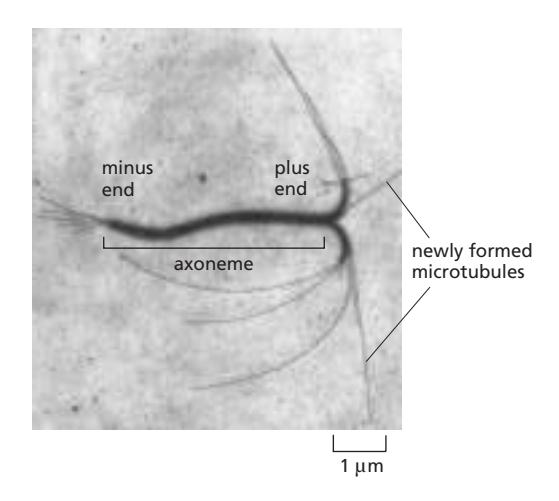 axoneme은 cilia나 Flagella의 중심축을 이루는 구조임. 9+2구조. 예외적으로 primary cilia는 9+0구조, 운동성 없고 안테나 역할만 함.
microtubule은 minus end 쪽(α-tubulin)보다 plus end 쪽(β-tubulin)에서 더 빠른 성장을 한다.
axoneme은 cilia나 Flagella의 중심축을 이루는 구조임. 9+2구조. 예외적으로 primary cilia는 9+0구조, 운동성 없고 안테나 역할만 함.