SNARE
개요
SNARE protein (SNAREs)은 vesicle transport에서 membrane fusion reaction을 catalyze하는 fusion protein이다.123 SNARE는 “SNAP receptor”의 약자로, 원래 NSF attachment protein (SNAP)과 상호작용하는 receptor로 발견되었다. 세부 메커니즘 : Vesicle Fusion by SNARE_MOC
SNARE의 종류와 분포
Classification
v-SNARE (vesicle-SNARE):4
- 주로 transport vesicle membrane에 위치
- Single polypeptide chain
- 대표적 예: Synaptobrevin (synaptic vesicle)
t-SNARE (target-SNARE):
- 주로 target membrane에 위치
- 보통 3개의 protein으로 구성
- 대표적 예: Syntaxin과 SNAP25 (plasma membrane)
Distribution
- Animal cell에 최소 35개의 서로 다른 SNARE
- 각 SNARE는 특정 organelle과 연관:
- Secretory pathway의 특정 단계
- Endocytic pathway의 특정 compartment
- Multiple SNARE가 한 membrane에 공존 가능
구조적 특징
Helical Domain
초기 상태:
- 대부분 unstructured
- Isolated 상태에서 defined structure 부재
활성 상태:
- v-SNARE와 t-SNARE interaction 시
- Four-helix bundle 형성
- 매우 안정한 구조
SNARE Complex
구성:
- v-SNARE: 1개의 α helix 기여
- t-SNARE: 3개의 α helix 기여
- Syntaxin: 1개 helix (transmembrane protein)
- SNAP25: 2개 helix (peripheral membrane protein, fatty acyl chain으로 anchor)
특징:
- Four parallel α helices가 coiled coil로 intertwine
- Extremely stable structure
- High energy of formation
Trans-SNARE Complex Formation
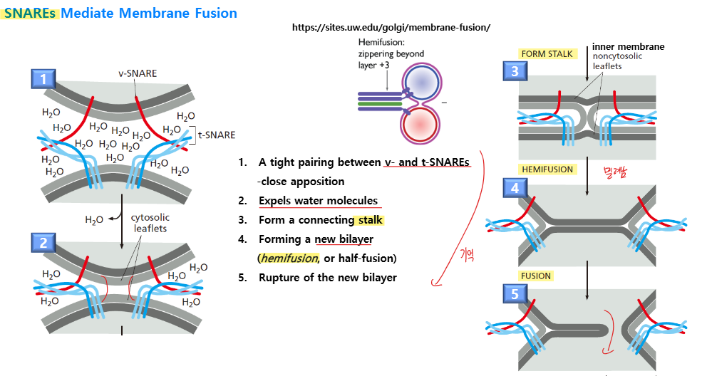
Zippering Mechanism
-
Initial Contact:
- v-SNARE와 t-SNARE의 N-terminal region 상호작용 시작
- Loose association
-
Progressive Zippering:
- N-terminus에서 C-terminus 방향으로 zippering
- Helical domain이 점진적으로 organize
- Membrane이 점점 더 가까워짐
-
Complete Assembly:
- Full four-helix bundle 완성
- 두 membrane이 매우 근접 (< 1.5 nm)
- Membrane fusion 준비 완료
Energy Release
Assembly process의 energetics:
- Unstructured helix → highly stable bundle
- 매우 energetically favorable
- Released energy가 다음을 drive:
SNARE Specificity
Pairing Specificity
SNARE pairing은 매우 특이적:
- 특정 v-SNARE는 특정 t-SNARE와만 pair
- Complementary set의 SNARE만 stable complex 형성
Functional Test
Liposome fusion assay:
- v-SNARE 포함 liposome 준비
- t-SNARE 포함 liposome 준비
- 혼합
- 결과:
- Matching pair: Efficient membrane fusion
- Non-matching pair: No fusion
Biological Significance
Specificity는 두 가지 level의 vesicle targeting 제공:
- Rab protein: Initial recognition과 tethering
- SNARE: Final recognition과 fusion
이중 체계로 매우 높은 targeting accuracy 달성.
Synaptic Vesicle의 특수한 SNARE
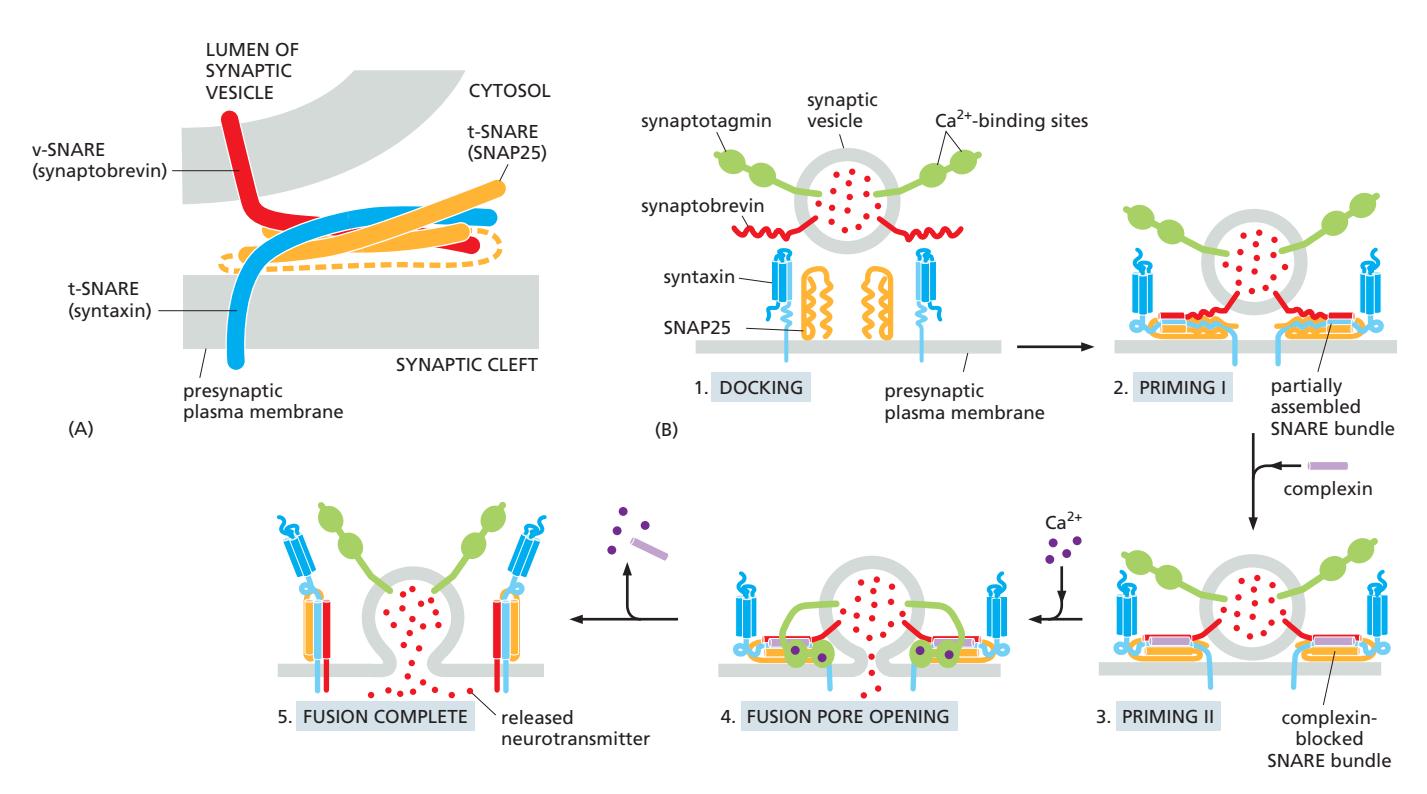
Synaptic vesicle fusion은 특수한 SNARE set 사용:
Component
- v-SNARE: Synaptobrevin (transmembrane protein)
- t-SNARE:
- Syntaxin (transmembrane protein, 1 helix)
- SNAP25 (peripheral membrane protein, 2 helices)
Regulation
일반적인 SNARE와 달리 추가 조절:
- Synaptotagmin: Ca²⁺ sensor
- Complexin: SNARE complex를 metastable state로 freeze
작동 메커니즘:
- SNARE partially assemble → primed state
- Complexin이 premature fusion 방지
- Ca²⁺ influx → Synaptotagmin 활성화
- Complexin release
- SNARE fully zipper → rapid fusion
- Neurotransmitter release (millisecond scale)
이는 regulated exocytosis의 특수한 형태.
Viral Fusion Protein
일부 virus는 자체 fusion protein 사용:
특징
- SNARE-mediated fusion과 달리 한쪽 membrane의 protein만 필요
- Virus envelope의 fusion protein
- Host cell membrane에는 특별한 fusion protein 불필요
Mechanism
- Appropriate environment에서 viral fusion protein unfurl
- Partially hydrophobic patch를 host membrane에 insert
- Fusion protein compaction
- 두 membrane을 가까이 bring
- Membrane fusion drive
예시
- HIV: Plasma membrane과 fusion
- Influenza virus: Endosome에서 low pH로 활성화
- SARS-CoV-2: Host protease cleavage로 활성화
Viral fusion은 SNARE와 유사한 원리지만 다른 molecular detail을 사용.
관련 내용
Footnotes
-
2022 중간 26번 — SNARE의 5단계(initial contact, progressive zippering, membrane fusion, NSF disassembly, recycling) 이해가 정답 근거로 활용됨. ↩
-
2023 중간 8번 — ①번 선지: Lipid bilayer 두 막의 세포질 면에서 만들어진 막에 새로운 bilayer가 만들어진다 (맞음, v-SNARE와 t-SNARE의 trans-SNARE complex가 membrane apposition·lipid merger를 drive, 정답); ②번 선지: 융합에 관한 SNARE 복합체(V-&T-SNARE)는 분리 후 그대로 보존된다 (틀림, NSF/α-SNAP에 의해 cis-SNARE complex가 disassembly된 후 재활용); ③번 선지: Hemifusion 단계에서 합쳐지는 bilayer의 한 층이 남아 필요 없어지면 분리된다 (틀림, hemifusion 단계에서 두 막의 outer leaflet만 먼저 fusion); ④번 선지: Stalk 형성 단계에서 lipid bilayer 두 막의 세포질 면의 인지질이 섞여 stalk를 만든다 (맞음, cytoplasmic face의 outer leaflet이 stalk 형성); ⑤번 선지: 소포와 target 막사이의 작동자가 먼저 연결될 때만 SNARE가 pairing 할 수 있다 (틀림, SNARE pairing 자체가 fusion을 drive하며 tethering 후에 가능). ↩ ↩2 ↩3
-
2023 중간 ver2 36번 — SNARE 5단계 서술형 문항; trans-SNARE complex 형성부터 NSF 분해 및 재활용까지 순서대로 서술해야 함. ↩
-
2021 중간 미상C번 — v-SNARE의 역할과 Rab effector(tethering protein, motor protein 등)에 대한 이해가 선지 근거로 활용됨. ↩
-
2025 중간 24번 — ①번 선지: 세포질 면의 막에서의 인지질이 초기 stalk를 만든다 (맞음, cytoplasmic face/outer leaflet의 인지질이 먼저 stalk 형성, 정답); ②번 선지: 세포질쪽에서 만들어진 막에 새로운 bilayer가 만들어진다 (틀림, 새 bilayer는 lumen 쪽에 형성); ③번 선지: Hemifusion 단계에서 합쳐지는 bilayer의 lumenal 막은 융합되지 않고 diaphragm이 형성된다 (맞음, hemifusion에서는 outer leaflet만 융합, inner/lumenal leaflet이 hemifusion diaphragm 형성); ④번 선지: 융합에 관한 SNARE 복합체 (V-&T-SNARE)는 분리되지 않고 보존된다 (틀림, NSF에 의해 분리 후 재활용); ⑤번 선지: Hemifusion 단계에서 합쳐지는 bilayer의 rupture를 통해 pore가 만들어지고 세포질 내에서 소포의 내용물이 희석된다 (틀림, pore 형성 후 내용물은 target compartment lumen으로 방출). ↩ ↩2